Advanced 3D Implant Diagnostics
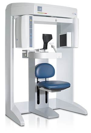
I-Cat CT Scan
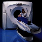
CT Scan machines (also known as CT Scan machines) have been used in the field of medicine for quite some time. The adjacent photo represents a typical configuration of a CT Scan device.
Advanced 3D Implant Diagnostics with a Cone Beam X-ray Machine
With the latest 3D x-ray or Cone Beam CT (CBCT) scanning technology, Dr. Brant is able to digitally diagnose dental problems and plan surgical procedures using high resolution 3-D scans of your mouth and teeth. Dr. Brant likes to make as many decisions before any incisions are made. Using the cone beam scanner is the only way to help with the pre-operative decision making process. Dr. Brant’s commitment to proper diagnosis before treatment is rendered led him to be the first dentist in Suffolk County to obtain an in-office Cone Beam CT scanner.
What is CBCT imaging?
CBCT is a type of X-ray equipment we use when regular x-rays are not sufficient. It can create 3-D images of your teeth, jawbones, soft tissues and nerve pathways using a single scan.
During the imaging process, a cone-shaped X-ray beam rotates around the patient’s head, producing up to 200 2-D images. Using specialized computer software, these images are then converted into a 3-D image, which can help us fully diagnose, and then treat, the problem.
Cone Beam Computed Tomography (CBCT) uses a special type of x-ray machine that can generate more information than a traditional x-ray. This machine is capable of producing high resolution 2D images or full 3D views of the mouth and sinuses that can give the doctor a better look at your teeth and other areas that might be causing you issues.
Cone Beam CT technology is not the same as conventional CT technology that you may have heard of. Cone Beam CT uses a cone shaped x-ray beam that moves around the patient, taking thousands of x-rays in a single rotation, that are then stitched together using software, which provides a panoramic, or 3D, view of the patients mouth. The images that the machine produces are similar to those of a conventional CT scan, but without the high radiation and need to go to an outside radiology center.
Benefits of CBCT imaging
First, the images can be shown to the prospective patient so a clear understanding can be gained by the patient about their own condition and how it will be treated. This “show and tell” session can be very interesting and instructive to some patients.
There are several benefits to using CBCT imaging, making it the preferred imaging method for many dental conditions.
It gives us a better idea of what’s going on inside your mouth. By using CBCT imaging, we get accurate measurements and a variety of views and angles, which makes for a more complete evaluation.
It can image bone and soft tissue at the same time. Unlike a typical dental x-ray, CBCT images provide information on your teeth, bones, and soft tissue in a single scan.
It’s quick! The scan typically takes between 20 – 40 seconds for a full mouth X-ray, and less than 10 seconds for a scan of a specific area.
A lower dose of radiation is used. There is much less radiation used with a CBCT scan than with a regular medical grade CT scan.
The machine itself is small. Unlike those scary x-ray machines you may see in your typical doctor’s office, CBCT scanners are actually very compact.
What Does Cone Beam CT Help With?
While x-rays provide our dentists with a good look at what’s going on in your mouth, CBCT scans offer us a much higher resolution look and a better understanding of what is happening and what needs to be done in order to treat the diagnosis.
There are many different uses for CBCT scans. Dr. Brant uses the Cone beam scanner to evaluate:
- the sinuses
- nerve canals
- jawbones and nasal cavity
- detecting and measuring jaw tumors
- Root canal problems
- getting a better look at bone structure and tooth orientation
- surgical planning for impacted teeth
- determining more accurate placement of dental implants
The following is a list of top reasons Dr. Brant will use the Cone Beam scanner.
- Illustrate available bone for implant placement. The relative health and maturity of the bone is demonstrated very well on the scan. In addition, the width and height of the bone of available bone is displayed very clearly. Computerized rulers can be used to accurately determine these dimensions.
- If the relative health and maturity of the bone is not known before the surgery, the implant surgeon may expose the bone only to find out that the bone is not ready or not adequate for the implant to be placed. No patient wants to have the area “opened up” for no good reason. A pre-op scan helps prevent this problem.
- The scan can be loaded into special dental implant computer software programs like Simplant or Galileos which allows the dental implant surgery to be performed on the computer. The placement of the dental implants in a virtual fashion allows Dr. Brant to choose the appropriate dental implant manufacturer for the case. Then we can choose the diameter and length of the anticipated dental implant.
- If the dimensions of the bone are not known beforehand, then the practitioner is guessing about what size implant he or she will need to place. If they guess wrong and don’t have the correct implant on hand to place, then the choice is to abort the surgery or place an inappropriately sized dental implant in the site. Neither instance is good news if you are the patient. Being able to load the Cone Beam scan data into advanced software like Simplant or Galileos saves the patient from this clinical outcome.
- Help extract teeth by showing how the roots exist in the bone which leads to less trauma at the time of extraction
- Predict what grafting is needed style and materials
- Predict likelihood of placing a dental implant at the time of extraction
- Show infection in bone not seen on regular dental x rays
- 3-d position and location of vital structures
- Show a fracture
- Show resorption
- What type of sinus lift procedure can be employed
- Relative health of the sinus
- Illustrate tooth positions for orthodontic purposes
Scanners in Medicine
A CT or CAT scan is the term used to describe a radiologic test known as Computerized Tomography or Computerized Axial Tomography.
The CT scanner has a doughnut shaped appearance that takes pictures of cross-sections of any part of the body or the entire body. These cross-sections are called slices and displayed digitally, either on a computer monitor or through digital photography.
Slices generated from this type of apparatus are approximately 1 millimeter thick (1000 microns). This type of tomography produces useable imagery about 60% of the time with magnification of the images ranging from 60 to 70 percent.
Adaptations in Dentistry
Periodontists and oral surgeons dinvolved with bone and tissue grafting of the maxilla or mandible started using CT Scan technology quite some time ago.
Over time however, committed dental surgeons discovered limitations that prevented acquiring all of the digital data desired that could promote a higher degree of accuracy. Radiation factors, thickness of the digital slices and magnification issues were felt to interfere with certain types of intricate dental surgeries.
Innovations leading to Virtual Dentistry
New CT Scan devices have been developed that have leap frogged the original CAT Scan innovation.
Cone Beam types of scanning devices have been developed that require little more than standing or sitting in a chair. The scanning device revolves around the patient, in much the same fashion as a Panoramic Xray machine commonly used by dentists.
Our I-CAT 3-D Imaging equipment, pictured here, enables Dr. Brant to produce precision scans with advanced levels of accuracy. (Click here to watch a short animated video demonstrating the convenience of this new technology).
Radiation is reduced by a significant… 95% amount. The Cone Beam Slices are a fraction of what Medical Scanners produce. A Cone Beam slice is 150 microns …. versus 1000 microns (one millimeter). Resolution is 500 percent greater.
This new CAT Scan standard now enables experienced dental surgeons to perform reconstructive dentistry and tissue grafting procedures with stunning accuracy.
Patients seeking additional information or details about specialized treatment sessions or appointments are invited to contact Dr. Brant directly via our on-line Ask The Dentist form during or after usual business hours.
Call: 631.584.4395
Dr. Brant can provide examples of how executives from a variety of industries and institutions are provided with a custom utilization of our services to deliver treatment outcomes for nearly any kind of time schedule.